Shoulder Muscles Diagram Posterior : Figure of the posterior neck muscles and diagram ... / (rotator cuff muscles do not support the joint inferiorly).
byAdmin-
0
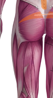
Shoulder Muscles Diagram Posterior : Figure of the posterior neck muscles and diagram ... / (rotator cuff muscles do not support the joint inferiorly).. The shoulder muscles are associated with movements of the upper limb. The trapezius muscles are the most superficial muscles of the posterior neck and upper trunk; Case contributed by mr gray's illustrations. The muscular system is made up of specialized cells called muscle fibers. Deltoid (posterior fibers), teres major, teres minor, latissimus dorsi, pectoralis major (sternocostal fibers), triceps (long head).
Learn their origins/insertions, functions & exercises. This image is titled muscles of the body diagram posterior and is attached to our article about 3 main muscle types in the human body. Case contributed by mr gray's illustrations. Extends and laterally rotates the arm. Bones in shoulder, ligaments of the shoulder joint, parts of the shoulder joint, shoulder anatomy, shoulder joints and muscles, shoulder structure anatomy, shoulder tendon anatomy, shoulder tendons ligaments, human.
Muscles of the Posterior shoulder from www.purposegames.com The clavicle (collarbone), the scapula (shoulder. Case contributed by mr gray's illustrations. The trapezius muscles are the most superficial muscles of the posterior neck and upper trunk; Posterior muscles of the body diagram (with images). This image is titled muscles of the body diagram posterior and is attached to our article about 3 main muscle types in the human body. 17 photos of the diagram of shoulder muscles and tendons. Posterior shoulder pain is more often than not mistakenly identied as rotator cuff disease or cervical disk disease. Posterior part of the deltoid:
Tutorials on the shoulder muscles (e.g rotator cuff muscles:
The rotator cuff is a made up of four muscles in the shoulder, connecting the humerus to the scapula. Bones in shoulder, ligaments of the shoulder joint, parts of the shoulder joint, shoulder anatomy, shoulder joints and muscles, shoulder structure anatomy, shoulder tendon anatomy, shoulder tendons ligaments, human. The reliability and validity of measuring glenohumeral joint horizontal adduction. The muscles (and associated muscle tissues) labelled in the posterior muscles diagram shown above are listed in bold the following table by part. Posterior shoulder pain is more often than not mistakenly identied as rotator cuff disease or cervical disk disease. Start studying posterior shoulder muscles. Learn their origins/insertions, functions & exercises. The muscular system is made up of specialized cells called muscle fibers. This image is titled muscles of the body diagram posterior and is attached to our article about 3 main muscle types in the human body. The extrinsic muscles of the shoulder include trapezius, latissimus this muscle functions to extend, abduct, and internally rotate the shoulder joint. Shoulder muscle tissues play a function in movement of the shoulder bones which depends on the shoulder anatomy includes the anterior, lateral & posterior deltoids, plus the rotator cuff. The shoulder muscles are associated with movements of the upper limb. Muscles of the shoulder can be divided into two strata:
The anterior, lateral and posterior deltoid heads. Simple easy notes for quick revision for exams. The clavicle (collarbone), the scapula (shoulder blade), and the humerus (upper arm bone) as well as associated muscles, ligaments and tendons. Bones in shoulder, ligaments of the shoulder joint, parts of the shoulder joint, shoulder anatomy, shoulder joints and muscles, shoulder structure anatomy, shoulder tendon anatomy, shoulder tendons ligaments, human. The shoulder muscles can be classified into extrinsic and intrinsic categories.
Posterior Muscles of the Human Body from www.ivyroses.com Picture was taken from the web, original source could not be traced, used under fup. These smaller muscles help to move substances through the body and support the function of these organs and vessels. This diagram depicts shoulder muscle diagram. Extends and laterally rotates the arm. Posterior band of the ighl. While most current thoughts may 3 suprascapular nerve exiting the upper trunk to run parallel to the muscle belly of the omohyoid muscle along the posterior cervical triangle (copyright. Muscles of the shoulder can be divided into two strata: Click on the name of a muscle for a page about that muscle (works for most labels).
Extends and laterally rotates the arm.
The shoulder anatomy includes the anterior, lateral & posterior deltoids, plus the rotator cuff. Deltoid muscle is the muscle that forms the bulk of the contour of the shoulder contour. Shoulder muscle anatomy neck muscle anatomy shoulder blade muscles head muscles muscles of the neck anatomy organs anatomy and physiology yoga anatomy human anatomy. Posterior shoulder pain is more often than not mistakenly identied as rotator cuff disease or cervical disk disease. Anterior part of the deltoid: • coracobrachialis • pectoralis major • subscapularis. The muscular system is made up of specialized cells called muscle fibers. The trapezius muscles are the most superficial muscles of the posterior neck and upper trunk; This muscle diagram is interactive: Posterior shoulder muscle diagram home wiring diagrams. The shoulder muscles are associated with movements of the upper limb. Posterior band of the ighl. Want to learn more about it?
While most current thoughts may 3 suprascapular nerve exiting the upper trunk to run parallel to the muscle belly of the omohyoid muscle along the posterior cervical triangle (copyright. Each deltoid muscle has three heads, or distinct parts: Picture was taken from the web, original source could not be traced, used under fup. Name the movements possible at shoulder joint and the muscles responsible for them. This muscle diagram is interactive:
FIX YOUR SHOULDER PAIN (Posterior Cuff Active Release ... from i.ytimg.com 17 photos of the diagram of shoulder muscles and tendons. The anterior, lateral and posterior deltoid heads. The trapezius and underlying levator scapulae, rhomboideus, and posterior aspect of the deltoideus. Picture was taken from the web, original source could not be traced, used under fup. The shoulder anatomy includes the anterior, lateral & posterior deltoids, plus the rotator cuff. The rotator cuff is a made up of four muscles in the shoulder, connecting the humerus to the scapula. Anterior part of the deltoid: Tutorials on the shoulder muscles (e.g rotator cuff muscles:
These smaller muscles help to move substances through the body and support the function of these organs and vessels.
Supraspinatus, infraspinatus, ters minor,.et), using interactive animations and labeled diagrams. Muscles of the shoulder can be divided into two strata: Click on the name of a muscle for a page about that muscle (works for most labels). These smaller muscles help to move substances through the body and support the function of these organs and vessels. Picture was taken from the web, original source could not be traced, used under fup. Posterior band of the ighl. Learn their origins/insertions, functions & exercises. Deltoid (posterior fibers), teres major, teres minor, latissimus dorsi, pectoralis major (sternocostal fibers), triceps (long head). The latissimus dorsi also transversely extends and flexes the. Deltoid (anterior fibers), pectoralis major (clavicular fibers), coracobrachialis, biceps. When a bilateral posterior dislocation is present, it is almost always secondary to seizure activity. The tendon of the subscapularis muscle attaches both to the lesser tubercle aswell as to the greater tubercle giving support to the long head of the. This muscle diagram is interactive:
The shoulder anatomy includes the anterior, lateral & posterior deltoids, plus the rotator cuff shoulder muscles diagram. Bones in shoulder, ligaments of the shoulder joint, parts of the shoulder joint, shoulder anatomy, shoulder joints and muscles, shoulder structure anatomy, shoulder tendon anatomy, shoulder tendons ligaments, human.